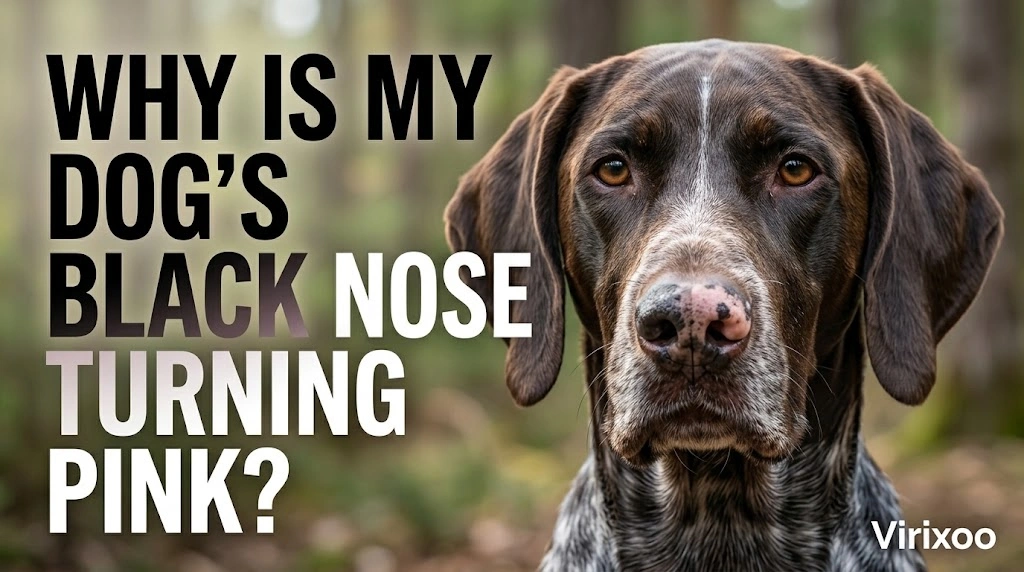
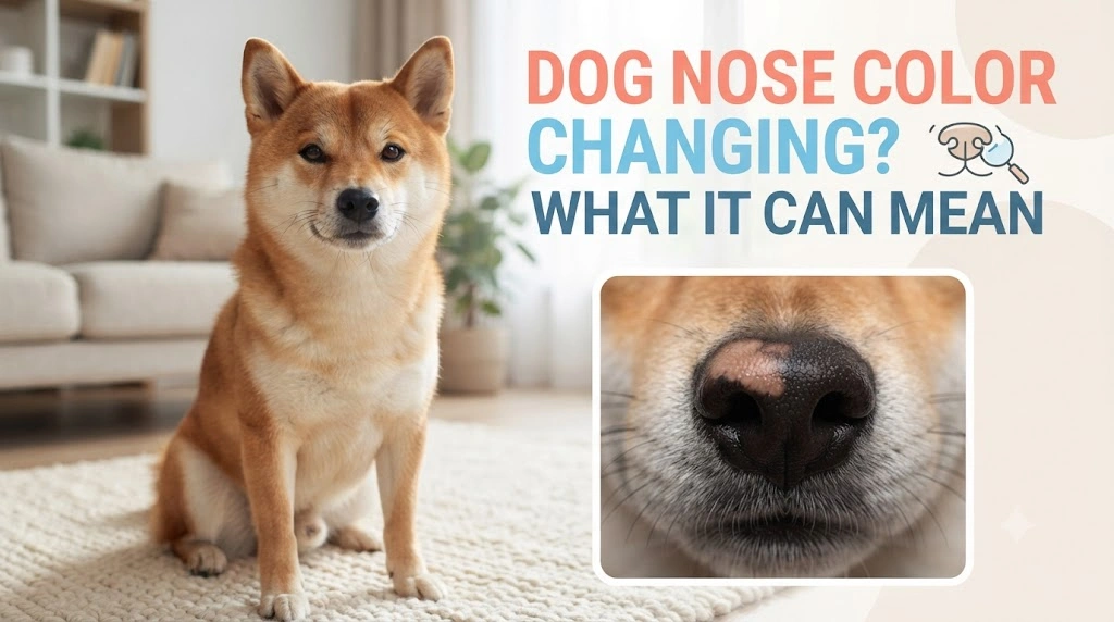
A dog’s nose can become lighter, darker, patchy, or seasonally faded for reasons that range from harmless pigment variation to inflammation, injury, immune-mediated disease, or other skin problems. Color alone rarely tells you how serious a change is. The more useful distinction is whether the nose is simply changing pigment while remaining smooth and comfortable, or whether color loss comes with crusting, sores, bleeding, swelling, pain, or lesions elsewhere.
Quick answer
If your dog’s nose gradually becomes lighter but remains smooth, normally textured, comfortable, and free of sores, the change may be benign. Take a clear photo in consistent lighting and monitor it. Contact a veterinarian when pigment loss is new and progressive, the surface becomes crusted or ulcerated, there is bleeding, swelling, discharge, pain, or similar lesions appear around the lips, eyes, or skin.
Normal nose pigment varies
Not every healthy dog has a uniformly black nose. Some dogs naturally have brown, liver, pink, mottled, or partially pigmented noses based on genetics. Puppies can also undergo pigment changes as they mature. The meaningful question is whether your dog’s nose looks different from its own established baseline.
Photos taken several months apart in natural light are often more useful than comparing your dog with another breed. Camera filters and indoor lighting can exaggerate color differences.
Seasonal fading
Some dogs develop a lighter nose during colder or lower-light seasons, often called seasonal or “snow” nose. The central portion of a previously dark nose may fade to brown or pinkish tones and darken again later. A seasonally lighter nose that remains otherwise healthy is different from a depigmented area that is raw, painful, cracked, or spreading with skin lesions.
Seasonal timing is a clue, not a diagnosis, because several conditions can alter pigment.
Friction and repeated rubbing
A dog that repeatedly rubs the nose against a crate, fence, carpet, or ground can damage the surface. Friction tends to produce irritation, abrasions, hair loss on nearby skin, or crusting rather than a neat pigment change alone. Identify and remove the mechanical trigger while protecting the area from further trauma.
Inflammation and skin disease
Inflammatory skin conditions can affect the nose and the junction where normal skin meets the nasal surface. Signs may include redness, scaling, crusts, erosions, loss of normal texture, or pigment change. Infection can develop secondarily when the skin barrier is damaged.
Because several diseases look similar in photographs, veterinary examination—and sometimes additional testing—is needed when lesions persist.
Immune-mediated conditions
Certain immune-mediated diseases can cause depigmentation, crusting, ulcers, or changes around the nose and other facial areas. These conditions cannot be diagnosed from color alone. Distribution of lesions, overall health, sun exposure, and sometimes laboratory or skin testing help the veterinarian determine the cause.
What about vitiligo?
Vitiligo can cause pigment loss in skin and hair in some dogs without making the area painful or inflamed. Other causes of depigmentation can look similar at first, so veterinary assessment is sensible when pigment loss is new, extensive, or progressive. Do not assume every smooth pink patch is vitiligo.
Sun exposure matters when pigment is lost
Less-pigmented skin may be more vulnerable to sunlight. If your veterinarian confirms a condition that causes depigmentation, ask which sun-protection strategy is safe. Do not apply human sunscreen to the nose without guidance because some products contain ingredients that may be inappropriate if licked.
Dryness is not the same as pigment loss
A dog’s nose can feel drier or wetter at different times of day without being ill. Color change is a separate observation. A dry but normally colored nose after sleep is different from progressive fading with crusts or ulcers. Nose moisture alone is not a reliable fever test.
A home comparison checklist
| What to check | More reassuring | More concerning |
|---|---|---|
| Timing | Gradual or seasonal | Sudden or rapidly progressive |
| Surface | Smooth and intact | Crusted, ulcerated, bleeding |
| Comfort | No rubbing or pain | Persistent rubbing or sensitivity |
| Distribution | Isolated pigment change | Lesions around lips, eyes, or other skin |
| General health | Normal appetite and energy | Lethargy, weight loss, or other illness |
Should you put moisturizer on the nose?
Do not automatically apply human creams, petroleum products, essential oils, or medicated balms. Dogs lick their noses, and a product considered safe for human skin may not be suitable when swallowed. More importantly, covering crusts or ulcers can delay diagnosis. Ask your veterinarian what, if anything, should be applied.
When color change follows an injury
A scrape or superficial wound can heal with temporary or permanent pigment alteration. If you know the dog recently injured the nose and the area is healing normally, photograph progress. Wounds that remain open, drain, swell, bleed repeatedly, or become more painful should be examined.
Could a bowl be involved?
Repeated contact with a rough or damaged bowl can irritate skin around the muzzle. Use clean, food-safe bowls in good condition and wash them regularly. However, switching bowls should not be treated as a cure for progressive depigmentation, crusting, or ulceration.
Texture is often more important than shade
A nose that changes from dark to light but keeps its usual texture is different from one that becomes smooth, eroded, thickened, cracked, or crusted. Texture change suggests the tissue itself is being affected, not merely the pigment.
Check the rest of the face and body
Look at the skin around the lips, eyelids, ears, and paw pads without manipulating sore areas. If pigment loss or crusting appears in several locations, tell the veterinarian. Also report hair loss, itching, joint pain, appetite changes, or lethargy because the nose may be part of a broader skin or immune problem.
Take useful progress photos
Use the same room or natural-light location, similar distance, and no filter. Include one close photo and one showing the whole face. Date the images. This helps distinguish a stable pigment variation from an expanding lesion and provides more reliable information than differently lit pictures.
When to call the veterinarian
Arrange an appointment if pigment loss is spreading, persistent, or accompanied by scaling, cracking, sores, bleeding, swelling, nasal discharge, pain, or changes around the eyes and lips. Seek prompt care for major facial swelling, severe bleeding, significant trauma, breathing difficulty, or sudden severe illness.
Bottom line
Nose color can change without disease, especially when genetics or seasonal fading is involved. What turns a pigment change into a medical concern is damage to the surface, pain, crusting, ulceration, bleeding, rapid progression, or additional skin and systemic signs. Photograph the change consistently, avoid unprescribed topical products, and let a veterinarian evaluate persistent or inflamed lesions.
Why change over time matters
Pigment changes are often gradual, and memory is unreliable. A monthly photograph in similar natural light can show whether a light patch is stable, seasonal, or steadily expanding. If the surface becomes ulcerated, crusted, painful, or bloody, do not wait for several comparison photos before seeking veterinary advice.
How to decide whether to monitor or call
Use your dog’s normal baseline as the reference. A mild, familiar pattern in a comfortable dog is very different from a new or rapidly changing problem. With dog nose color changes, contact a veterinary professional sooner when pain, weakness, bleeding, vomiting, breathing changes, appetite loss, or rapid progression appears. Online guidance can organize what you observe, but it cannot replace an examination when your dog is becoming unwell.
When you do call, give a simple timeline: when the change began, whether it is getting better or worse, what other symptoms are present, and what medications or recent exposures may be relevant. Clear information helps the veterinary team triage the situation efficiently.
Why change over time matters
Pigment changes are often gradual, and memory is unreliable. A monthly photograph in similar natural light can show whether a light patch is stable, seasonal, or steadily expanding. If the surface becomes ulcerated, crusted, painful, or bloody, do not wait for several comparison photos before seeking veterinary advice.
What if hair around the nose changes too?
Loss of pigment in nearby hair or skin, hair loss, scaling, or lesions around the eyes and lips provides additional clues. A cosmetic pigment change limited to the nose is different from a broader facial skin disorder. Report any itching, rubbing, joint pain, appetite loss, or lethargy because these signs may help the veterinarian decide whether testing should extend beyond the nose itself.
Why texture often matters more than color
A smooth pink patch can be very different from a crusted or eroded pink patch. When the normal surface architecture disappears, the issue is no longer purely pigment. Cracking, bleeding, thickening, and ulceration indicate tissue damage and deserve veterinary evaluation even if the dog seems otherwise comfortable.
What the veterinarian may examine
The veterinarian may look closely at the nose, lips, eyelids, ears, paw pads, and other skin. Depending on the pattern, testing can include cytology, culture, blood work, or biopsy. Not every dog needs extensive testing; the distribution and surface changes guide the next step.
Before you decide the change is cosmetic
Look beyond color. A smooth, painless patch that changes slowly is very different from a nose that is crusted, cracked, swollen, bleeding, or losing its normal texture. Also check whether similar changes are appearing around the lips, eyes, or other skin. If the dog feels unwell or the lesion is progressing, veterinary assessment is more appropriate than trying new creams, supplements, or home remedies.
Why Texture Matters as Much as Color
A smooth light patch can be very different from a crusted, eroded, thickened, or bleeding patch. When the normal surface texture disappears, the issue is no longer purely pigment. Photograph the nose in consistent natural light and look for changes around the lips, eyelids, ears, or paw pads as well.
Monitoring is reasonable only when the dog feels well, the surface is intact, and the change is gradual. Spreading lesions, pain, crusting, ulceration, bleeding, or other illness signs deserve veterinary evaluation.